


A new type of scanner shows promise in generating never-before-seen brain tumour images and is expected to help in treatment of glioblastoma patients.
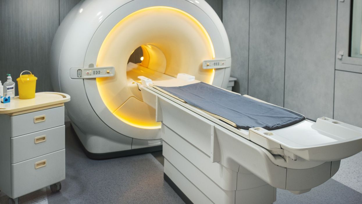
The Field Cycling Imaging (FCI) scanner developed at the University of Aberdeen will provide more precise images than MRI scans.
It can detect tumours without having to inject dye into the body, which can cause allergic reactions and kidney damage in some patients.
This technology has already been found to be effective in detecting tumours in breast tissue and brain damage in stroke patients.
Researchers at the University of Aberdeen and NHS Grampian have been awarded £350,000 funding by the Scottish government to carry out a trial on a group of 18 glioblastoma patients.
FCI scanners can work at low and ultra-low magnetic fields and provide images of affected organs in ways that were previously not possible.
Researchers hope FCI will be able to tell the difference between tumour growth and progression, and "pseudo-progression," which looks like a tumour but is not cancerous.
This facility is not available in MRI scans.
MRI scanners were invented at the University of Aberdeen 50 years ago.
Glioblastoma is the most common type of brain tumour, with more than 3,000 new patients in the UK diagnosed each year.
Half of all glioblastoma patients die within 15 months of diagnosis, even after getting treated.